There are a number of errors that can happen when an organism develops from an embryo to an adult. A single missing cell or a cell out of place can have catastrophic results for the matured individual. Even a slightly altered rate of cell division at such an early stage is more likely to cause eventual death than anything else, with less dire results including severe malformation of limbs and body structure.
Many different kinds of genetic disorders get their start this way and it has proven difficult to follow the developmental steps that lead to the consequent problems. Because when it comes to complicated genetic problems that can’t just be fixed by genetic editing, the only other option is to understand how the cell types divided and organized themselves within the body. But following the original group of cells into the trillions they ultimately become is no easy task.
Many Attempts, Few Successess
The lineage relationships between early cells, their adult divisions, and even the cells of descendant offspring, are all important factors in measuring and understanding such disorders. But methods like single cell RNA (scRNA) sequencing are highly limited and often can only separate out cell types, but are unable to differentiate the developmental origins of said differentiated cells.
For a time, fluorescent proteins attached to genes were used in order to follow cellular development, but the optical fidelity of such techniques don’t have the resolution needed to isolate the amount of cells that are required. It works when limited to only a handful of cells, but any more than that go beyond the ability to trace optically. That research then led to other options that each sought to expand the amount of recognized cells during development. These included things like viral barcoding, somatic mutations, microsatellite repeats, and more, but they all ran into the problem of only following lineage, but not being able to differentiate out cell types.
In order for any of this to work, both are a necessity, the capability to follow the differentiation of embryonic cells to maturity and to classify the cell types each such cell turns into once multiplied. Without the latter, the former can’t be connected to the particular genetic disorders being studied, which may often affect specific cell formats in the body. So scientists had to find a method that was able to do both or, more likely, a combination of methods that would jointly allow both to be accomplished.
CRISPR-Mediated Genetic Scarring
Researchers at Germany’s Max Delbrück Center for Molecular Medicine took up that task and aimed to make a tool that would be able to flawlessly complete both requirements. And, as might be expected, the primary device they co-opted for the task was CRISPR-Cas9.
It’s already well known that when CRISPR is used to cut the DNA strands, especially when cutting both strands, this is likely to introduce new nucleotide additions that interfere with the function of a gene. In a way, it is a form of point mutation as directed by human guidance. The researchers thought to use this in order to make a specific series of marks, what they referred to as genetic scarring, that would be passed on to all later cells and on to new generations.
They decided to use a zebrafish as a representative model organism line that can be reasonably compared to humans and also chose a specific breed that already has an inserted transgene for red fluorescent protein (RFP). This, as you might guess, causes the zebrafish to glow red under specific lighting. The transgene used was integrated in a broad number of copies, from 16 to 32, spread across the entire genome.
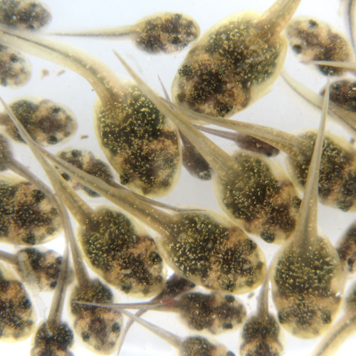
The scientists injected the Cas9 with a guide RNA into a one cell zebrafish embryo targeting said RFP transgene. If successful, the fish would fail to glow when inspected, showing that the Cas9 had done its job. Then, the zebrafish were allowed to grow from single celled embryos to the larvae stage. They were then dismantled and had cells from various organ tissues placed into single cell cultures for analysis.
Following The Cellular Timeline
Single cell RNA sequencing (scRNA-Seq) was used on around 70,000 individual cells after the five day growth period. The unique cell lines were differentiated and then each type had its RFP transgene sequenced. It was found, as desired, that the Cas9 had produced hundreds of unique genetic scars in the gene, depending on the cell type, even though it has only been targeting a single location in the transgene.
Further analysis showed that certain forms of scarring sequences were more likely to occur, possibly through some form of micro-homology repair that didn’t manage to completely fix the altered sequence. The Cas9 was shown to remain active in the differentiating cells for up to 10 hours after the single cell began division, meaning it was allowed to make all those unique scars in each new cell over time, creating the plethora of special scarring that match each cell type.
If this approach is then combined with scRNA-Seq during the early embryonic differentiation period, the unique scars on the RFP (or any other gene decided on for other organisms) can be recorded for each line at the beginning and then any cells found with that specific scar in the adult organism will be known to have come from that particular earlier embryonic cell. However, they did notice one complication in the overall process.
The number of scars in some cells varied significantly, from as many as 5 to as few as 2 depending on the cell type. This means that the overall amount of lineage information was lower on the latter group of cells and they determined that the efficiency of scarring detection depends on the cell type, likely related to both cell size and strength of the particular promoter sequence for the transgene in that cell. But all highly expressed scars were found to be detectable overall, even in specific cell types, so this is unlikely to be a major concern when using this method.
A Recording of Biological Development
The researchers have opted to call this new tool for cell type lineage measurements LINNAEUS, which stands for lineage tracing by nuclease-activated editing of ubiquitous sequences. They hope that it will serve as a basic framework for future experimentation into cell lineages and that it will allow proper tracing of them and molecular recording of important cell events during growth from the embryo stage.
Photo CCs: Early embryonic development of Encarsia pergandiella. Nature as art or vice versa? from Wikimedia Commons