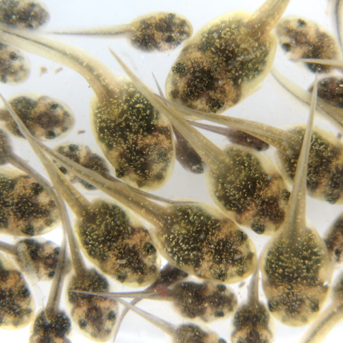
The field of embryology continues to be one with more mysteries than answers. Since it is difficult in many countries around the world to work directly with human embryos, animal models are often the best we can do. To a certain extent, stem cells allow us to at least observe the earliest stages of multicellular formation. We’ve discussed those sorts of things several times before.
But to go further along in development to the blastocyst stage where cell differentiation truly begins in earnest and to see how the extracellular membrane involving the placenta is created, there currently is no better option than actual embryos. And there is increasing evidence that mouse animal models do not actually match the same genetic activation and resulting regulatory effects that humans do.
Embryo Gene Targeting
To showcase this even further, a new study conducted by the Francis Crick Institute and in collaboration with many other universities and organizations around the world gained permission from the UK Human Fertilisation and Embryology Authority to work with 58 human embryos obtained from discarded in-vitro fertilization (IVF) treatments. The purpose was to determine the function and effect of a particular regulatory gene known to be involved in embryo development.
The same gene would be turned off in both the human embryos and in mouse embryos, to see what impact a lack of the resulting protein would have and whether that result would match between species. The gene in question they targeted is called POU5F1 and is a gene that begins being actively transcribed and translated into proteins starting around the 4 to 8 cell division stage. The protein is a developmental regulator known as OCT4 and only begins appearing at detectable levels at the 8 cell stage.
Due to its usage so early in the embryonic cycle, inactivation of it would be expected to have a major impact on later cell division and differentiation.
Accurate Guides
To identify and target the gene, an optimized CRISPR-Cas9 system was used, with four specialized single guide RNAs (sgRNA) made for this purpose. Each one targeted a different exon in the POU5F1 gene. Exons are the coding part of a gene that are actively made into later protein amino acid sequences. Exons are usually surrounded by and have in between them introns, which are non-coding DNA segments that are spliced out of the later messenger RNA.
As was expected, the most effective guide RNA proved to be the one targeting the highly conserved portion of the gene responsible for proper folding and expression of the resulting protein. It caused heavy loss of OCT4 expression, with only 15.6% of the cells created having any detectable amounts. The other guide RNAs, assessed individually, had around 40-70% of their cells still express the protein.
Then, live embryo imaging was used to visualize the effect this genetic change was having on the interior cells of the blastocyst. The overall structure formed as normal, but the inner cell mass was found to lack proper cellular integrity, causing the entire blastocyst to eventually collapse inward.
The Mice Effects
Transcriptomics was then used to see what influence turning off POU5F1 has on the rest of the genome. What the researchers found was that genes involved in three other primary gene groups used in various preimplantation stages are downregulated in response to POU5F1 not being active. Combined, these appear to have the destructive result as just noted.
When compared to the outcome with mouse OCT4 deficient cells, the differences were striking. The most impressive guide RNA from before only managed a 54% success rate in mice, despite the gene being targeted being the same. The other guide RNAs had a success rate between 0 and 10%.
Also, it was discovered that OCT4 activation and usage appears to occur sooner in humans than in mice. And it didn’t appear that other genes took over the regulatory functions during these early stages in mice. Therefore, the genes involved at these cell division stages must be different in mice altogether, with POU5F1 not coming into play until later. Even when the right stages did start, mice retained functionality of several genes that are downregulated in human embryos.
Based on this, it appears that the OCT4 protein has just a different usage between humans and mice, making the latter not a suitable model for such research. It is likely that, in the future, human embryos may be the only proper option for research into how early cell differentiation occurs.
An Opening For Embryology
A positive thing to note in addition to what was already learned about OCT4’s regulatory involvement is that it also appears to have an effect on the cells that later form the placenta. Since placental development is one of the most mysterious areas of embryology currently, the researchers that conducted the study are excited to try further testing and develop a better understanding of this area of research.
Such a proof of concept on using CRISPR-Cas9 in conjunction with embryo regulatory genes should allow future scientists to further knowledge on embryo formation even more, improving the success rate of treatments like IVF and possibly allowing for better therapeutic development of stem cells.
Photo CCs: Mouse embryo from Wikimedia Commons


